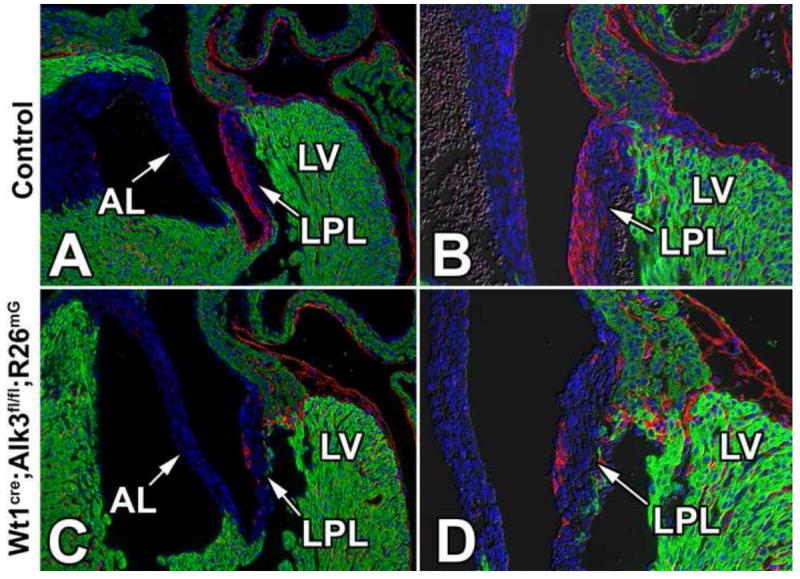
Figure 7. EPDCs fail to populate the parietal leaflets in Wt1cre;Alk3fl/fl;R26mG hearts.

Immunolabeling for eGFP (red), MF20 (green), and nuclei/DAPI (blue) in ED17 control (A,B) and Wt1cre;Alk3fl/fl;R26mG (C,D) specimens. Panels C and D show a sharp reduction of eGFP-labeled EPDCs in the left parietal leaflet (LPL) of the Wt1cre;Alk3fl/fl;R26mG mouse. AL, aortic leaflet; LPL, left parietal leaflet; LV, left ventricle.
