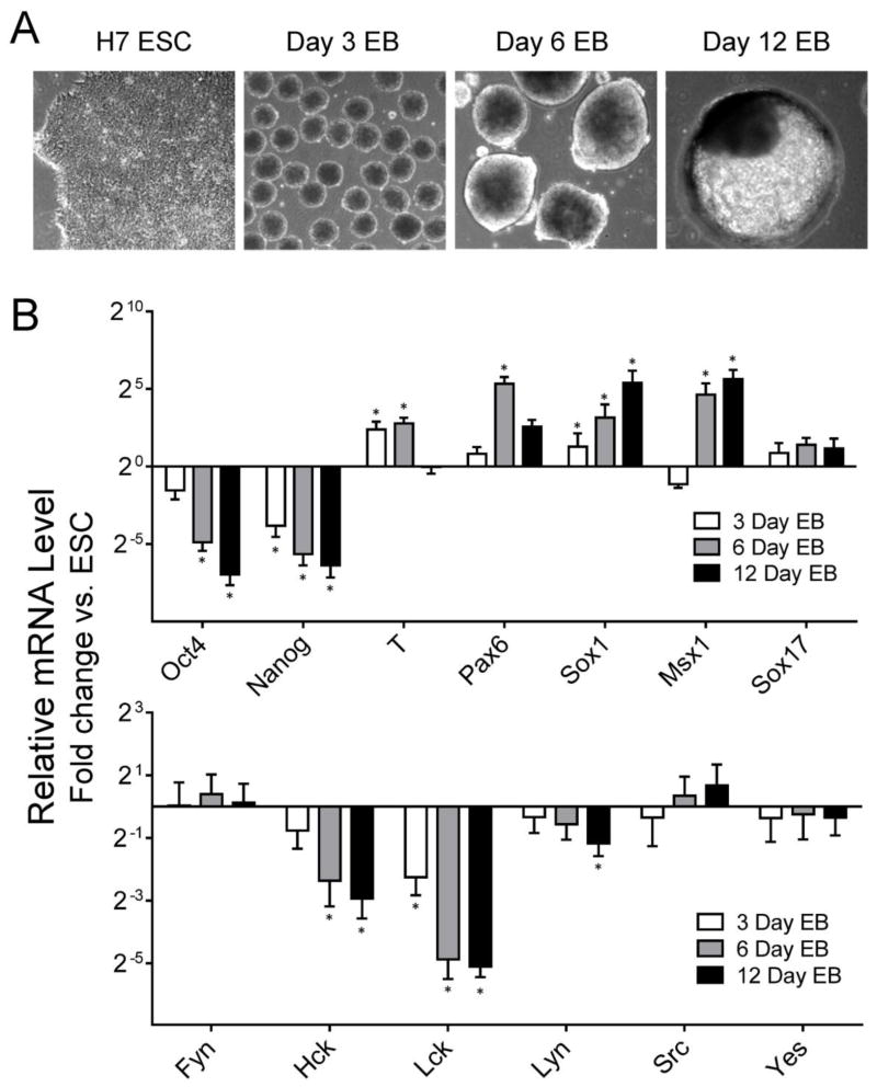
Figure 2. SFK expression during EB formation from H7 hES cells.
A) EB formation was initiated from H7 hES cells using Aggrewell plates as described under Materials and Methods. Representative images of H7 hES cells and EBs after 3, 6 and 12 days of culture later are shown; all images recorded at same magnification (100 ×). B) Total RNA was extracted from renewing hES cells and 3, 6, and 12-day EBs. Expression of self-renewal (Oct4, Nanog) and differentiation markers (T, mesoderm; Pax6, Sox1, Msx1, ectoderm; and Sox17, endoderm) as well as SFKs (Fyn, Hck, Lck, Lyn, c-Src, c-Yes) was determined by qPCR relative to control H7 hES cells maintained in mTeSR medium. Results are expressed as the average fold change relative to the undifferentiated hES cell population ± S.E.M. (n = 3; *p < 0.05; Pairwise Fixed Reallocation Randomization Test).

