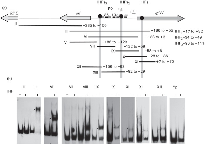
Fig. 5.
Binding of purified IHF heterodimer to the ygiW–qseBC promoter. (a) Schematic representation of the fdhE–ygiW intergenic region showing the putative binding sites for IHF. PCR fragments used for EMSA reactions are numbered from II to XIII, and the nucleotides contained by each fragment are indicated to the right, and numbered relative to the ygiW start codon. (b) PCR probes were incubated in the absence (−) and presence (+) of 4 µM IHF protein for 30 min at room temperature, and the DNA–protein complexes (indicated by an asterisk) were resolved in 6 % polyacrylamide gels. A 200 bp DNA fragment from Y. pseudotuberculosis psa (probe Yp) was used as a negative control.

