
Figure 6. Overexpression of NFATc1 in MC38Par cells increases tumor incidence and liver metastases.
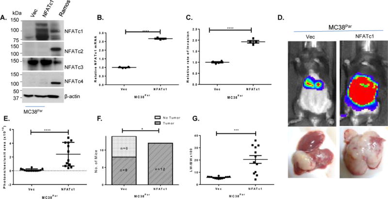
(A) Western Blot showing NFAT proteins in MC38Par cells transfected with either empty vector (VEC) or NFATc1, β-actin is used as a loading control and Ramos extract as positive control. (B) Analysis of NFATc1 mRNA in MC38Par cells shown in (A). (C) Rates of trans-endothelial invasion for MC38Par cells shown in (A–B). Individual replicate wells from a representative experiment are plotted with the mean and the standard error of the mean (bars and whiskers). (D) Representative mice from splenic metastasis model (n=12–14/group) using MC38Par cells shown in (A–D). (E) Bioluminescence of mice injected with MC38Par cells shown in (A–D) at 14th days. (F) Incidence of liver metastases for mice injected at day 14. (G) Liver weight to body weight ratio in mice injected with either MC38Par shown in (A–F) cells at day 14 post-injection. *p<0.02, *** p=0.0004, **** p<0.0001.