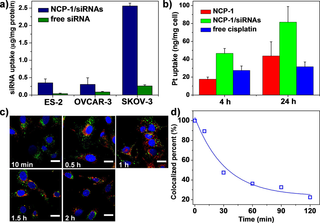
Figure 2.
(a) Cellular uptake amount of siRNA in ovarian cancer cells after incubating for 4 h (n=3). (b) Cellular uptake amount of Pt in SKOV-3 cells after incubating for 4 h and 24 h (n=3). (c) Time-dependent endosomal escape of NCP-1/siRNAs (TAMRA-labeled, red fluorescence) in SKOV-3 cells. Endosome/lysosome and nuclei were stained with Lysotracker Green and DAPI, respectively. Bar represented 20 µm. (d) Percent co-localization of siRNA and endosome/lysosome quantified by Image J based on (c).

