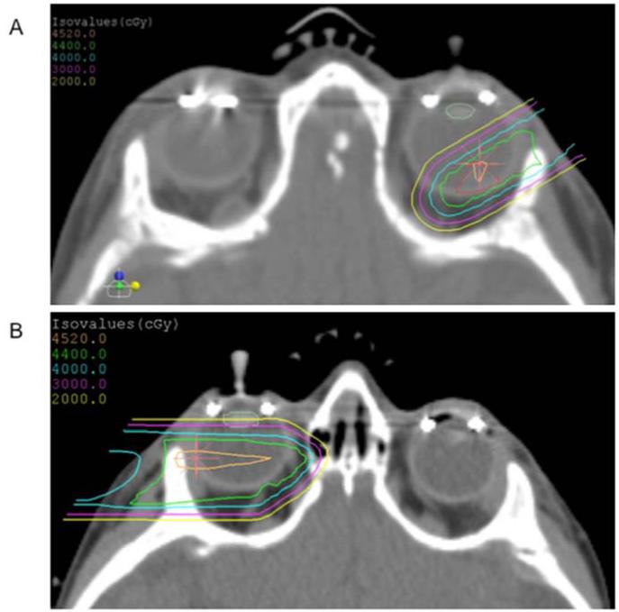
Figure 1.
Representative axial CT slice from two PRT plans. A. For small tumors without seeding, a single anterior oblique beam was used to minimize dose to the bony orbit (prescription dose 44 Gy[RBE]). The tumor is outlined in red, and the lens is outlined in pale green. B. For larger tumors, or if seeding was present, the posterior chamber was targeted with a single lateral beam (prescription dose 45 Gy[RBE]).

