
Figure 3. Paracellular flux routes vary along the crypt-villus.
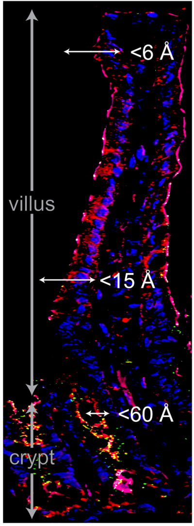
Fluorescence microscopy showing F-actin (red), claudin-2 (green), and nuclei (blue) in mouse jejunum. Note the increased claudin-2 expression in the crypt region. Tight junctions in the upper villus are permeable to small molecules (<6 Å), such as glucose, while channels with radii estimated to be 10-15 Å have been described in the lower part of the villus. Crypt tight junctions are permeable to macromolecules with radii up to 60 Å. Along with enhanced expression of claudin-2, which mediates paracellular Na+ and, possibly, water flux, this may promote net water secretion into the crypt space.