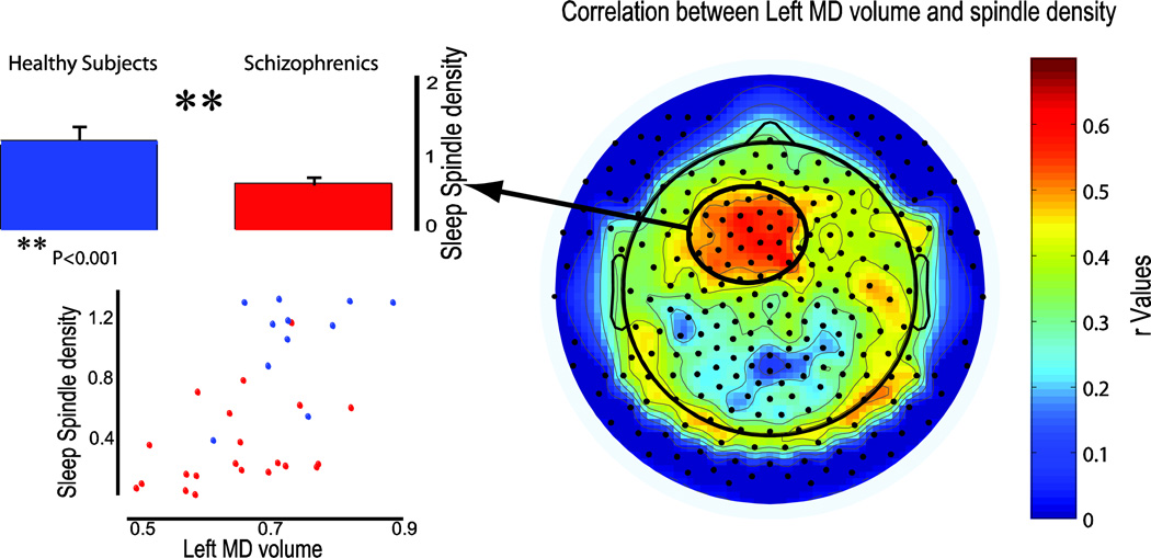
Figure 2. Left MD volume reduction in schizophrenia patients significantly correlated with sleep spindle density in a frontal cluster of electrodes.
This cluster (black-encircled red spot) was significantly reduced in schizophrenics compared to healthy controls (blue and red box plots, p<0.01). On the bottom left, single subject correlation between left MD volume and spindle number for schizophrenia patient (red) and healthy controls (blue).

