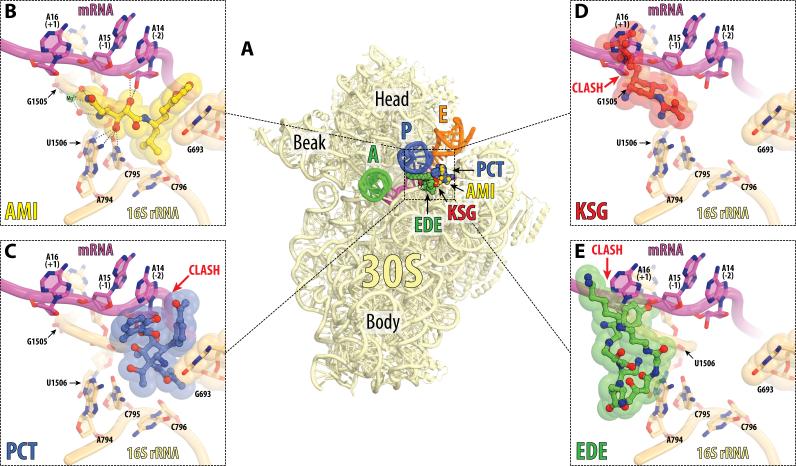
Figure 4. Antibiotics in the E site on the small ribosomal subunit.
(A) Overview of the superimposed binding sites of AMI (yellow), PCT (blue), KSG (red), and EDE (green) on the 30S subunit. The view and the coloring of 16S rRNA, mRNA and tRNAs are the same as in Figure 2A. AMI and PCT structures are from the current work, KSG is from PDB entry 1VS5 (Schuwirth et al., 2006), and EDE is from PDB entry 1I95 (Pioletti et al., 2001). All four structures were aligned based on h24 of the 16S rRNA (nucleotides 769-810). (B, C, D, E) Close-up views of the binding sites shown in (A) for AMI, PCT, KSG, and EDE, respectively. Steric clashes between antibiotics and parts of the ribosome are indicated by red arrows. Note, that AMI tethers mRNA to the 16S rRNA and does not clash with any parts of the ribosome, while PCT, KSG and EDE clash with mRNA. See also Figure S4 and Movie S2.

