Figure 1.
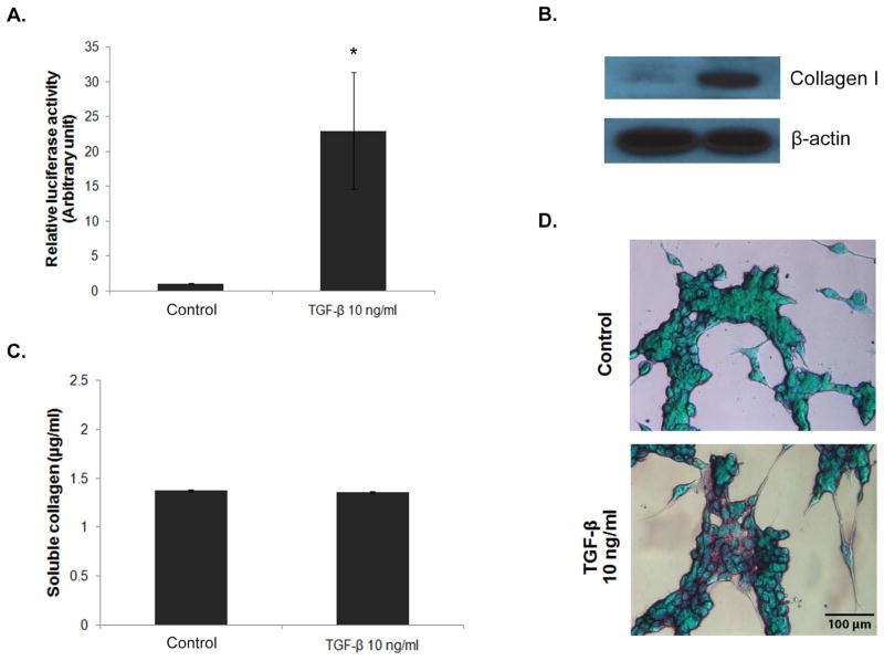
TGF-β-induced SMAD pathway and collagen production. Stable 3T3/NIH-SMAD-luciferase cells were generated from lentiviral transduction followed by puromycin selection. Cells were seeded into a 6-well plate at the density of 100,000 cells/well and incubated overnight. After a pre-incubation period of 6 hrs in serum-free medium, the cells were treated with 10 ng/ml of TGF-β1 for 16 hrs and analyzed for SMAD signaling activity and collagen production by (A) luciferase assay, (B) Western blot assay, (C) Picro-Sirius red assay, and (D) Sirius red/fast green staining. * = significant difference from control with P<0.05. Wild-type 3T3/NIH cells were used as a negative control for luciferase assay. Values obtained from the wild-type cells were subtracted from the test values before calculating relative luciferase activity.

