Figure 2.
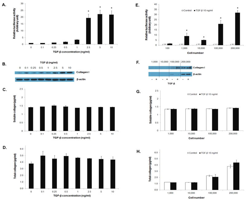
Comparison of detection sensitivity of various collagen assays. 3T3/NIH-SMAD-luciferase cells were seeded into a 6-well plate at the density of 100,000 cells/well and treated with TGF-β1 at different concentrations ranging from 0.1 to 10 ng/ml for 16 hrs. SMAD signaling activity and collagen type I expression were determined by (A) luciferase assay and (B) Western blot assay. Soluble and total collagen content was measured by Picro-Sirius red assay (C and D). Cell number-dependent detection sensitivity was demonstrated by treating the reporter cells at different cell numbers with 10 ng/ml of TGF-β1 and measuring luciferase activity and collagen content (E to H). * = significant difference from control with P<0.05.

