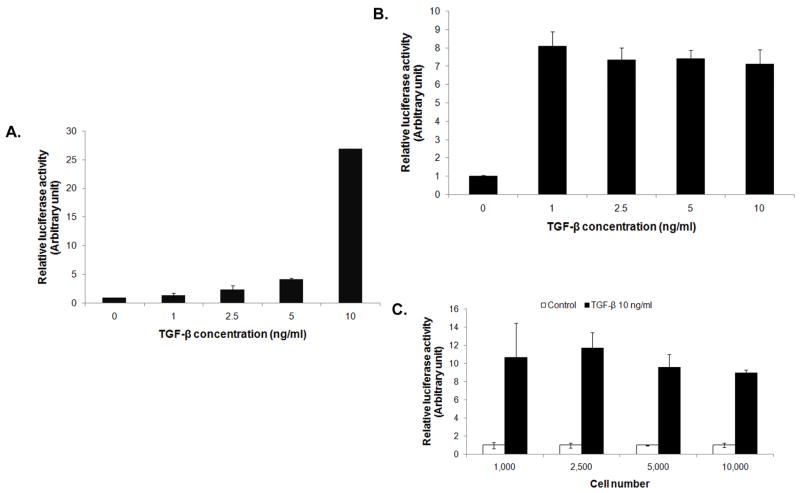
Figure 3.
Adaptability of 3T3/NIH-SMAD-luciferase reporter cells to different detection platforms. (A) Reporter cells were introduced into a single-channel microfluidic device and their luciferase activity in response to different concentrations of TGF-β1 was examined after 16 hrs using GloMax® 20/20 luminometer. (B) Induction of luciferase activity of the reporter cells by TGF-β1 in a 96-well format was evaluated. Reporter cells (10,000 cells/well) were treated with different concentrations of TGF-β1 and analyzed for luciferase activity using FLUOstar® OPTIMA plate reader. (C) Cells at different seeding densities were treated with 10 ng/ml of TGF-β1 and analyzed for luciferase activity.

