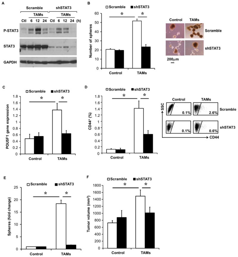
Figure 7. STAT3 signaling is critical for TAM-induced CSC expansion in HCC.
(A) Disrupted HepG2 STAT3 activation following TAM co-culture in STAT3 shRNA HepG2 cells. Scramble shRNA or STAT3 shRNA HepG2 cells were co-cultured with TAMs for indicated time (hours). Phospho-STAT3 and total STAT3 protein levels were assessed by western blotting. One of three representative experiments is shown.
(B) Scramble shRNA or STAT3 shRNA HepG2 cells were co-cultured with TAMs for 3 days, and then collected for sphere assay (n=5). One representative photomicrograph (10x magnification) is shown.
(C) Co-cultures with scramble shRNA or STAT3 shRNA HepG2 cells and TAMs were performed as in (B) and POU5F1 gene expression determined by qRT-PCR (n=5).
(D) Inhibition of TAM-mediated CD44+ subset expansion in vivo following STAT3 knockdown. Scramble shRNA or STAT-3 shRNA HepG2 cells were injected IP into NSG mice and six weeks later TAMs (30 million) were IP injected and CD44 subset determined by FACS three days later (n=5 mice/group and representative FACS).
(E) STAT3 knockdown blocks TAM-induced spheres. Experiment performed as in (D) and HCC tumor single cells were placed into sphere assays (n=9–10 tumors/group).
(F) Loss of TAM-mediated tumor-promoting effects in STAT3 knockdown HepG2 cells. Scramble shRNA or STAT-3 shRNA HepG2 cells (1000) co-cultured without or with TAMs were injected subcutaneously into NSG mice. Tumor volume at 42 days after cell implantation is shown (n=5 mice). One of three representative experiments is shown.
*p < 0.05. Data presented as mean±SEM.

