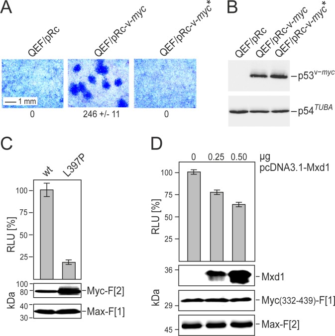
Fig.2. Correlation of Myc cell-transforming potential and Myc-Max interaction.
(A) Cell transforming potential of v-Myc and the dimerization-defective mutant v-Myc* (L397P). Quail embryo fibroblasts (QEF) were transfected with 6-μg aliquots of the plasmids pRc-v-myc, pRc-v-myc*, or with the empty pRc vector. Cells were kept under agar overlay for two weeks and then stained with eosin methylene blue. Foci were counted on 60-mm dishes (a section is shown; representative experiment of n=2, ± SD of triplicates). (B) Overexpressed HA-tagged v-Myc or v-Myc* proteins, and endogenous tubulin were analyzed by immunoblot analyses of QEF cell extracts prepared one day after transfection. (C) PPI of Myc-Max were quantified in Rluc-PCA experiments. Rluc-PCA signals were detected from chemically transformed QT6 cells transiently expressing Max-F[1]:v-Myc-F[2] (wt) or Max-F[1]:v-Myc*-F[2] (L397P) PCA pairs. Rluc-PCA tagged hybrid proteins were analyzed by immunoblot analysis. (D) SW480 cells stably expressing the RLuc-PCA pair Myc(332-439)-F[1]:Max-F[2] were subjected to bioluminescence analysis following transient expression of Mxd1 (HA-tagged). Increasing amounts of pcDNA3.1-Mxd1 vector DNA were transfected (representative of n=3; ± SD from triplicates). Expression of the PCA hybrid proteins and of Mxd1 was analyzed by immunoblotting.

