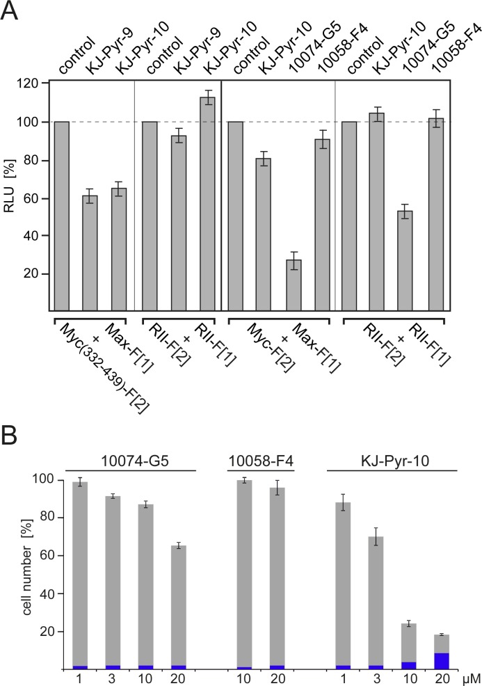
Fig.3. Effect of small-molecule Myc inhibitors on cellular PPI and cell proliferation.
(A) HEK293 cells were transiently transfected with the indicated Rluc-PCA Myc:Max (cf. Fig. 1B) or PKA-based RII:RII expression constructs. Bioluminescence was detected from control cells, or cells treated for 6 h with KJ-Pyr-9, KJ-Pyr-10, 10074-G5, or 10058-F4 (all at 20 μM) (± SEM from at least n=4 independent experiments). (B) Effect of Myc inhibitors on human cancer cell proliferation. Dose-dependent effects of the indicated Myc inhibitors on proliferation of the human T-cell leukemia cell line MOLT-4. Cells were exposed to the compounds for 72 h. Cell counts in percent of untreated control cell numbers were determined, and the fraction of non-viable cells (shown in blue) was quantified using a trypan blue assay (± SD of n=3 independent experiments).

