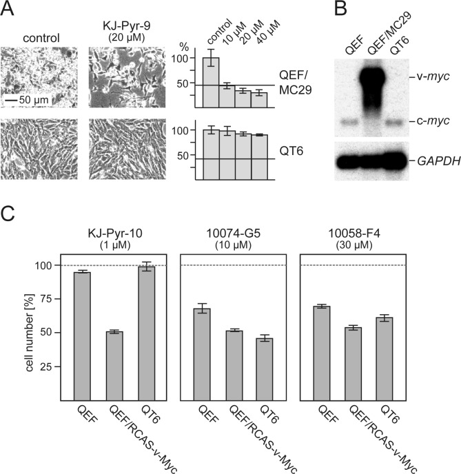
Fig.4. Efficacy and specificity of small-molecule Myc inhibitors.
(A) Cells from the v-myc-transformed quail cell line QEF/MC29 or the methylcholanthrene-transformed quail cell line QT6 were treated for 24 h with inhibitor compound KJ-Pyr-9 at the indicated concentrations. Then, cells were counted and microphotographs were taken. Average numbers of control cells were set to 100%. Horizontal bars indicate the numbers of cells initially seeded. (B) Northern analysis of RNAs from QEF/MC29, QT6, and normal QEF using a myc-specific probe. The positions of v-myc (MC29 genomic) and c-myc mRNAs are indicated. Hybridization with a GAPDH-specific probe was used as RNA loading control. (C) Normal QEF, QEF transformed with an RCAS-v-Myc construct, and QT6 cells were treated for 48 h with inhibitor compounds KJ-Pyr-10, 10074-G5, or 10058-F4 at the indicated concentrations. Cell counts were determined and the numbers of control cells were set to 100% (representative of n=3, ± SD from triplicates).

