
Figure 6. MUC1-C is necessary for self-renewal.
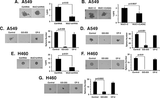
(A and B) Representative images are shown for the indicated A549 cells plated at 2000 cells/well and grown for 5 days in sphere culture (left). Bar represents 100 microns. The percentage SFE is expressed as the mean±SD of three determinations (right). (C) A549 cells were plated at 2000 cells/well in sphere culture and left untreated (Control) or treated with 5 μM GO-203 or CP-2 for 3 days. Representative images on day 5 are shown for the indicated A549 cells (left). The percentage SFE is expressed as the mean±SD of three determinations (right). (D) A549 cells were plated at 2000 cells/well and cultured for 5 days. The established spheres were then left untreated (Control) or treated with 5 μM GO-203 or CP-2 for 3 days. Representative images are shown for the indicated A549 cells (left). The percentage SFE is expressed as the mean±SD of three determinations (right). (E) Representative images are shown for the indicated H460 cells plated at 1500 cells/well and grown for 5 days in sphere culture (left). Bar represents 100 microns. The percentage SFE is expressed as the mean±SD of three determinations (right). (F) H460 cells were plated at 1500 cells/well in sphere culture and left untreated (Control) or treated with 5 μM GO-203 or CP-2 for 3 days. Representative images on day 5 are shown for the indicated H460 cells (left). The percentage SFE is expressed as the mean±SD of three determinations (right). (G) H460 cells were plated at 1500 cells/well and cultured for 5 days. The established spheres were then left untreated (Control) or treated with 5 μM GO-203 or CP-2 for 3 days. Representative images are shown for the indicated H460 cells (left). The percentage SFE is expressed as the mean±SD of three determinations (right).