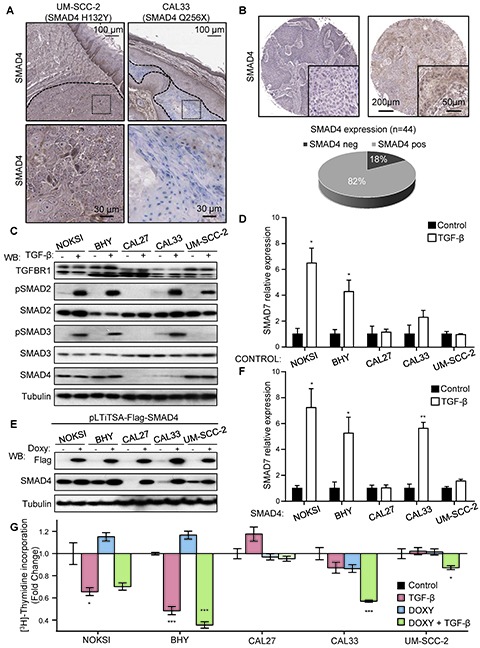
Figure 3. Aberrant TGF-β signaling in HNSCC and the OPC-22 panel.

(A) Sequencing data identified the presence of mutations in CAL33 and UM-SCC-2 as indicated. A representative SMAD4 staining on tumor xenografts with an antibody raised against the C-terminus of SMAD4 detected expression (brown) in UM-SCC-2 tumors and mouse stroma, while CAL33 tumors were negative. Tumor areas are delimited by a dashed line. Dotted area insets are shown at higher magnification in the corresponding lower panels. (B) Analysis of a cohort of 44 HNSCC cases stained for SMAD4. A representative negative (left) and positive (right) case is shown. Whole cohort quantification is shown in the lower panel. (C) Analysis of the TGF-β signaling in select HNSCC-derived lines. Cells were cultured under exponential growing conditions and then serum starved overnight. Cells were stimulated with vehicle (-) or 100 ng/ml TGF-β (+) for 45 minutes. Cells lysates were analyzed by Western blot for the proteins and phospho-proteins indicated in the figure. (D) Cells cultured as in C, were stimulated for 6h with vehicle (black bars) or 100 ng/ml TGF-β (white bars) and then RNA was extracted. SMAD7 expression was determined by qPCR. n=4, *, p≤0.05 for TGF-β different from Control. (E) A doxycycline inducible Flag-SMAD4 WT-IRES-GFP lentivirus was engineered and used to infect HNSCC as indicated. The percentage of SMAD4 WT expressing cells was determined to be over 70% in each case by GFP analysis. Cells cultured in the presence of 1 μg/ml doxycycline for 18h were lysed and analyzed by Western blot. (F) Cells in exponentially growing conditions were serum starved 12h in the presence of 1 μg/ml doxycycline and then stimulated for 6h with vehicle (black bars) or 100 ng/ml TGF-β (white bars). RNA was then extracted and SMAD7 expression levels determined by qPCR. n=4, *, p≤0.05, **, p≤0.01. (G) Cell proliferation assay by [3H]-thymidine incorporation. Exponentially growing cultures in the presence or absence of doxyclycline as indicated for 24h. Cells were then serum starved and treated with TGF-β while maintaining doxycycline treatment. [3H]-Thymidine (1μCi) was added to the cultures 4 h before the end of the treatment (total treatment time 24h). n=4, *, p≤0.05, ***, p≤0.001
