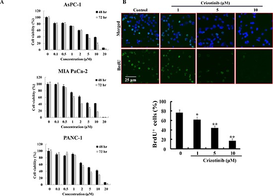
Figure 1. Effect of Crizotinib on the proliferation of human pancreatic cancer cells.

(A) Cell viability of Crizotinib was measured by MTT assay in AsPC-1, PANC-1, and MIA PaCa-2 cells at 48 hr or 72 hr. (B) BrdU staining was performed after PANC-1 cells were treated with or without various concentrations of Crizotinib for 6 hr (*p < 0.05 and **p < 0.01, compared to control). The upper panel represents a treatment with various concentrations of Crizotinib. The below panel shows the percent of BrdU-positive cells in several random fields of the BrdU staining images. All results are expressed as a percent cell proliferation relative to the control.
