
Figure 4.
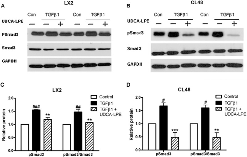
UDCA-LPE reduces TGF-β1-mediated phosphorylation of Smad3. (A,B) Representative Western blot analysis of pSmad3 and Smad3 in (A) LX2 hepatic stellate cells or (B) CL48 liver cells incubated with control medium, TGF-β1 (4 ng·mL−1) or TGF-β1 + 90 μM UDCA-LPE for 1 h. GAPDH was used as control for equal protein loading. (C,D) Graphical presentation of relative protein levels of pSmad3 and pSmad3/Smad3 ratio as estimated by densitometric analysis, values were relative to GAPDH and normalized to untreated controls, N = 3. #P < 0.05, ##P < 0.01, ###P < 0.001 versus Con; **P < 0.01, ***P < 0.001 versus TGF-β1.