
Figure 1. Morphological features of cells with slow waves filled with propidium iodide.
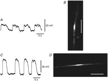
A, slow waves recorded from a cell in the longitudinal layer shown in B. The shape of the cell injected with propidium iodide for 5 min was digitally reconstructed on a confocal microscope (B). C, slow waves recorded from a cell in the circular layer shown in D. The shape of the cell injected with propidium iodide for 5 min was digitally reconstructed on a confocal microscope (D). Cells in B and D are of the general morphology of SMCs in the longitudinal and circular muscle layers. The scale bars in B and D represent 50 μm. The resting membrane potentials were: A, −46 mV; C, −64 mV. A and C were recorded from different tissues.