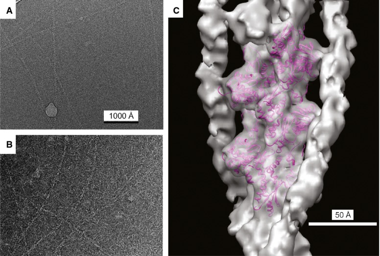
Figure 3. Three-dimensional reconstruction of F-actin with bound pacsin2.

A Electron cryo-micrograph of naked, unstained frozen/hydrated F-actin.
B Electron cryo-micrograph of unstained frozen/hydrated F-actin decorated with pacsin2. Magnification settings are same as in (A).
C The surface of the reconstructed volume is shown in gray, with an atomic model of F-actin 26 shown as magenta ribbons. The remaining density running along each actin strand is pacsin2.
