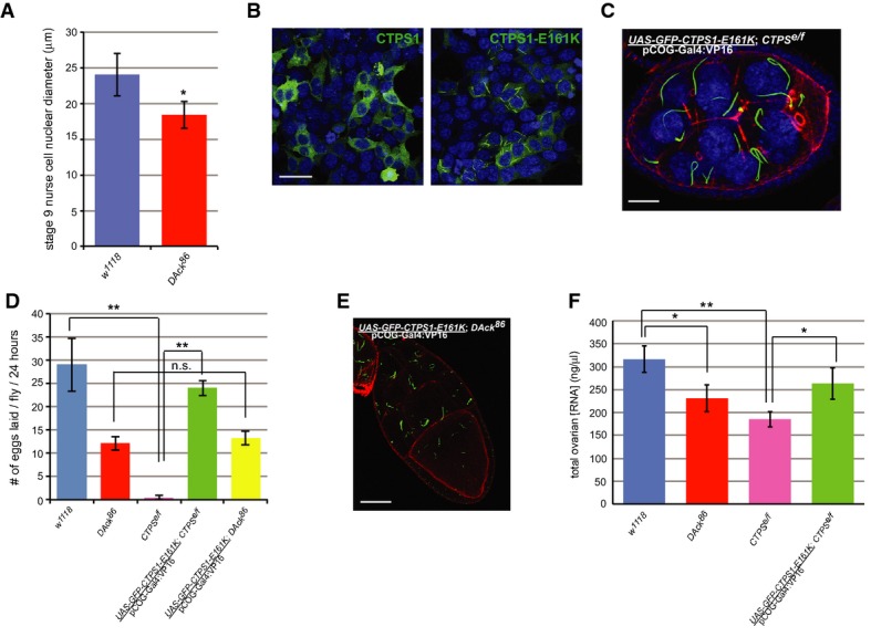
Figure 4. Catalytically active CTPS forms filaments.

A Defective endoreplication in DAck86 egg chambers. Diameters of nurse cell nuclei were measured from stage 9 egg chambers (n = 30) of each genotype. Shown is the average diameter with error bars indicating standard deviation. * denotes P-value < 0.05 as calculated by an unpaired, two-tailed Student’s t-test.
B Localization of constitutively active CTPS1. HEK293 cells transfected with constructs encoding GFP-tagged human CTPS1 or CTPS1-E161K were imaged by confocal microscopy. Scale bar, 50 μm.
C Localization of human GFP-CTPS1-E161K (green) in Drosophila ovarian germline cells. A three-dimensional confocal projection from a stage 8 egg chamber of the indicated genotype stained with rhodamine-phalloidin (red) and Draq5 (blue). Scale bar, 20 μm.
D Egg laying rates from flies of the indicated genotypes. The average rate from three independent experiments is shown. Error bars indicate standard deviation. ** denotes P-value < 0.005 as calculated by an unpaired, two-tailed Student’s t-test, and n.s. denotes not significantly different from w1118.
E Localization of human GFP-CTPS1-E161K (green) in Drosophila ovarian germline cells. A 0.4-μm confocal section from a stage 10 egg chamber of the indicated genotype stained with rhodamine-phalloidin (red) is shown. Scale bar, 40 μm.
F Quantitation of total ovarian RNA from flies of the indicated genotypes. Equal volumes of ovarian tissue were dissected from each genotype, and total RNA was isolated and measured. The average concentration from three independent experiments is shown. Error bars indicate standard deviation. * denotes P-value < 0.05, and ** denotes P-value < 0.005 as calculated by an unpaired, two-tailed Student’s t-test.
