
Figure 3.
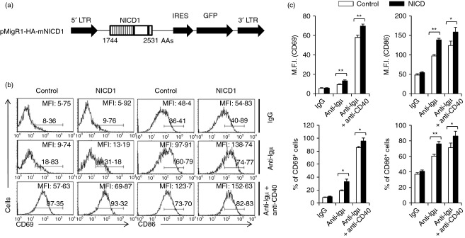
NICD1 expression in primary peripheral B cells increased B-cell activation. (a) Construction of mNICD1 expression retroviral vector. (b) B cells were isolated from C57BL/6 mice and the cells were infected with recombinant retroviruses for expression of mNICD1 and stimulated with either anti-µ F(ab)2 or anti-µ F(ab)2 plus anti-CD40 antibodies. (c) The summarized data from the flow cytometry analyses are shown (± SD) for mean fluorescence intensity and percentage of gated populations. Data are representative of three independent experiments. Data are representative of two independent experiments. Student’s t-test: *P < 0·05; **P < 0·01.