
Figure 5.
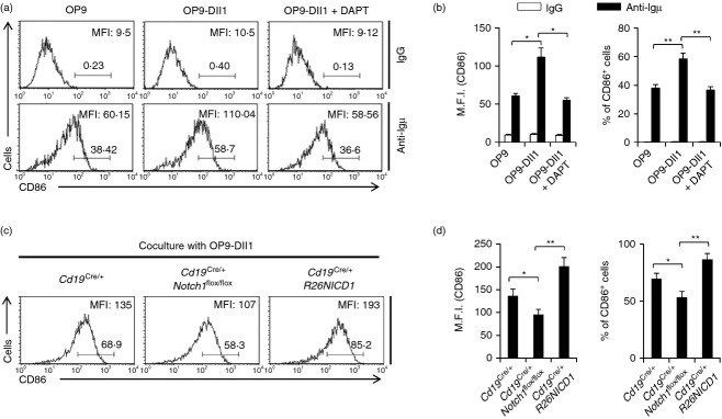
Notch1 deficiency abrogated Notch ligand-mediated increase in B-cell activation. (a) B cells were isolated from C57BL/6 mice and the cells were stimulated by anti-µ F(ab)2 antibodies with either OP9, OP9-Dll1 or OP9-Dll1 plus 50 µm of N-[N-(3,5-Difluorophenacetyl-L-alanyl)]-S-phenylglycine tert.butyl ester (DAPT) for 16 hr. The cells were stained with fluorochrome-conjugated anti-CD86 antibodies and analysed by flow cytometry. (b) The summarized data from the flow cytometry analyses are shown (± SD) for mean fluorescence intensity and percentage of gated populations. (c) B cells were isolated from the spleens of Cd19Cre/+, Cd19Cre/+ Notch1flox/flox and Cd19Cre/+ R26NICD1 mice and stimulated by anti-µ F(ab)2 antibodies with OP9-Dll1 for 16 hr. The cells were stained with fluorochrome-conjugated anti-CD86 antibodies and analysed by flow cytometry. (d) The summarized data from the flow cytometry analyses are shown (± SD) for mean fluorescence intensity and percentage of gated populations. Data are representative of two (a, b) or three (c, d) independent experiments. Student’s t-test: *P < 0·05; **P < 0·01.