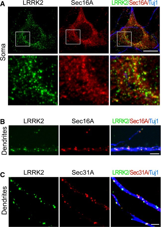
Figure 6. LRRK2 co-localizes with Sec16A proteins at the dERES of cultured hippocampal neurons.

A, B Representative images show LRRK2 (green) and Sec16A (red) staining in the soma (A) and dendrites (B) of Lrrk2+/+ hippocampal neurons at 14DIV. Neurons were marked by staining with an antibody (Tuj1) against βIII-tubulin (blue). Scale bar: 10 μm.
C Representative images show of LRRK2 (green) and Sec31A (red) in dendrites of hippocampal neurons at 14DIV. Neurons were visualized by staining with an antibody (Tuj1) against βIII-tubulin (blue). Scale bar: 10 μm.
