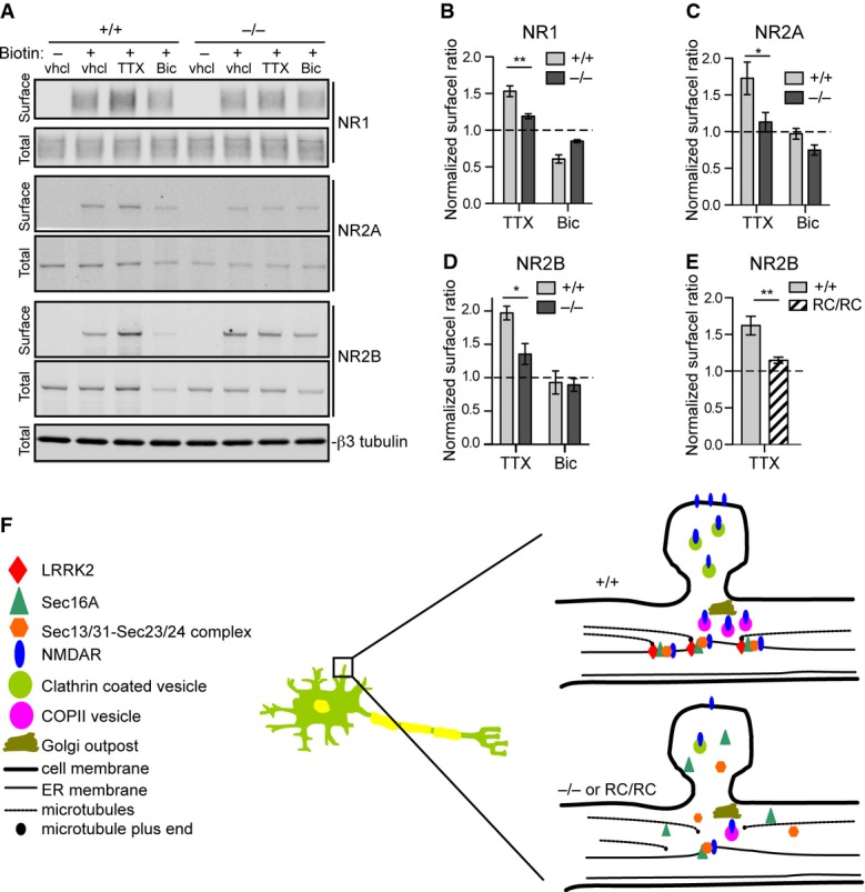
Figure 8. Impaired activity-dependent NMDA receptor trafficking in Lrrk2−/− and Lrrk2RC/RC neurons.

A Western blots show the levels of biotinylated cell surface and total NMDAR subunits NR1, NR2A, and NR2B in cultured Lrrk2+/+ and Lrrk2−/− cortical neurons (14DIV) treated with vehicle (vhcl), tetrodotoxin (TTX), and bicuculline (Bic), respectively. βIII-tubulin was used as the loading control for the lysate.
B–D Bar graphs show the ratio of NR1 (B), NR2A (C), and NR2B (D) at cell surface versus total proteins. The data from TTX and Bic-treated samples were normalized with vehicle-treated samples. Three and six independent experiments were performed on Lrrk2+/+ and Lrrk2−/− neurons, respectively. Two-way ANOVA shows significant interaction between genotype and TTX treatment for NR1 (F = 17.49, P = 0.0013), NR2A (F = 9.07, P = 0.027), and NR2B (F = 6.68, P = 0.022). *P < 0.05. **P < 0.01.
E Bar graph shows the ratio of NR2B at cell surface versus total proteins from vehicle and TTX-treated cultured Lrrk2+/+ and Lrrk2RC/RC cortical neurons (14DIV). The data from TTX-treated samples were normalized with vehicle-treated samples. Three independent experiments were performed on Lrrk2+/+ and Lrrk2RC/RC neurons. Two-way ANOVA shows significant interaction between genotype and TTX treatment for NR2B (F = 12.37, P = 0.0079). **P < 0.01.
F Schematic diagram illustrates a working model on how activity-dependent surface trafficking of NMDARs is impaired by redistribution of Sec16A in Lrrk2−/− and Lrrk2RC/RC neurons. LRRK2 may anchor the dERES near the dendritic spines through interaction with the dynamic ends of microtubules that are enriched at the base of dendritic spines (Jaworski et al, 2009), and facilitates the cargo transport from dERES to the dendritic spines in response to strong neuron activation.
Source data are available online for this figure.
