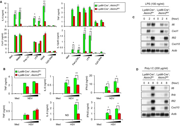
Figure 1. TLR-ligand- and virus infection-induced cytokine production and gene expression in Akirin2-deficient macrophages.
A, B Peritoneal macrophages from LysM-Cre+; Akirin2fl/+ and LysM-Cre+; Akirin2fl/fl mice were stimulated with various TLR ligands including Malp2 (1, 10 ng/ml), poly I:C (10, 100 μg/ml), LPS (10, 100 ng/ml), R848 (10 ng/ml) and CpG DNA (0.1, 1 μM) (A) or infected with HSV and NDV (MOI 1, 5) (B) for 24 h. Then, IL-6, TNF, CXCL-1, IL-12p40, and IFN-β concentrations in the culture supernatants were determined by ELISA. Error bars indicate mean ± SD. Results are representative of at least three independent experiments. Statistical significance was determined using the Student’s t-test. *P < 0.05; **P < 0.01.
C, D Peritoneal macrophages from LysM-Cre+; Akirin2fl/+ and LysM-Cre+; Akirin2fl/fl mice were stimulated with 100 ng/ml LPS (C) or poly I:C (200 μg/ml) (D) for indicated time period, and the total RNA extracted was subjected to Northern blot analysis for the expression of Il6, Ifnb, Cxcl1, Ifit2, Cxcl10, and Actb.

