
Figure 1. Trisomic and tetrasomic human cell lines show defects in protein folding.
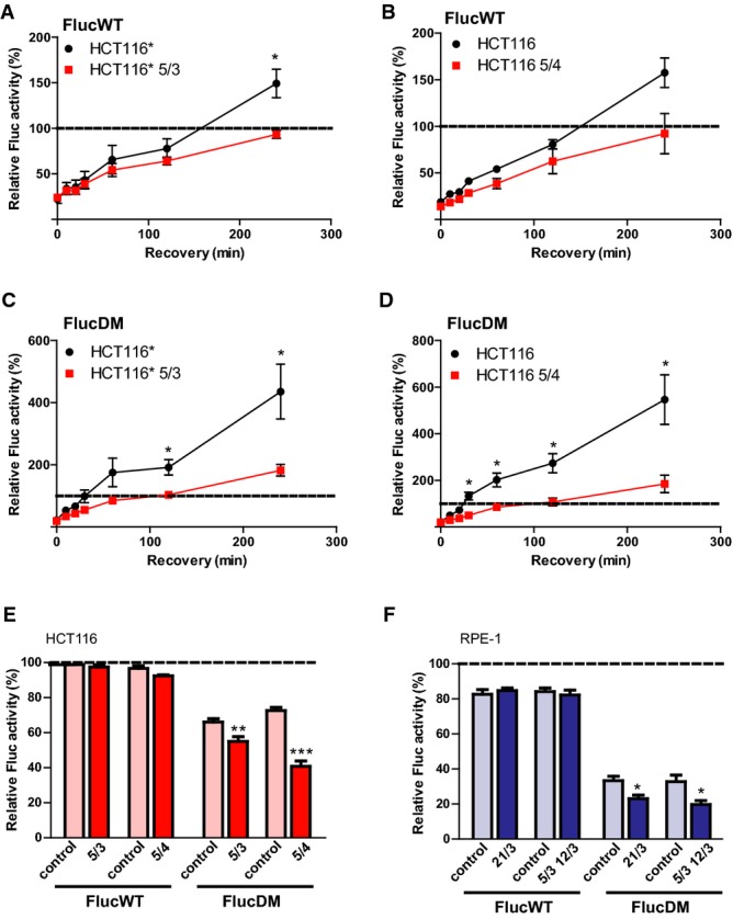
A–D Refolding of the sensor proteins upon heat shock in control cells and in respective aneuploids. HCT116 and HCT116* stably expressing histone H2B-GFP and their aneuploid derivatives were transfected either with FlucWT-mCherry (A, B) or FlucDM-mCherry (C, D) and subjected to heat stress for 2 h at 43°C. Controls were maintained at 37°C. Luminescence readings were taken immediately from heat-stressed cells (0 min) and at indicated time points after recovery at 37°C. The luminescence values of control cells maintained at 37°C were set to 100% (indicated by dotted line).
E, F Refolding of the sensor proteins upon HSP90 inhibition in control cells and in respective aneuploids. FlucWT-mCherry or FlucDM-mCherry was expressed in parental and aneuploid HCT116 or HCT116* (E) and RPE-1 or RPE-1* (F) cell lines for 36 h. Cells were then incubated with either solvent control (DMSO) or 50 nM 17-AAG for 8 h followed by measurement of luminescent activity. The depicted values show the percentage of luminescence in cells treated with 17-AAG relative to DMSO-treated cells (which were set to 100%).
Data information: All plots show the means of at least three independent experiments ± SEM. *P < 0.05; **P < 0.01; ***P < 0.001; non-parametric t-test.