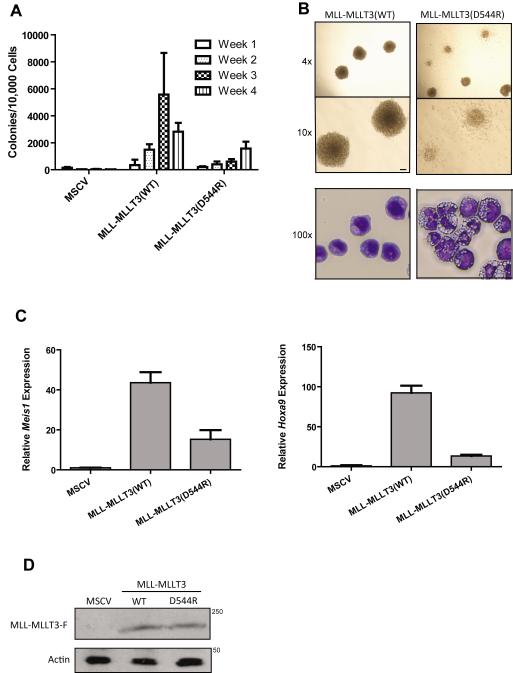
Figure 2. MLLT3-AFF1 interaction is required for MLL-MLLT3-induced transformation.
a) Cells expressing MLL-MLLT3(WT) efficiently re-plated, whereas cells expressing MLL-MLLT3(D544R) showed a statistically significant reduction in colony-forming ability (p<0.02). Colony assays were conducted in duplicate and repeated ten times. b) Top panel, MLL-MLLT3(D544R) expressing cells exhibited more diffuse colony morphology as compared to the dense, compact colonies formed by MLL-MLLT3(WT) cells; bottom panel, cytospin and staining with Wright-Giemsa indicate that MLL-MLLT3(WT)-expressing cells exhibit blast-like morphology, whereas cells expressing MLL-MLLT3(D544R) appear differentiated. c) Expression of MLL-MLLT3 target gene Meis1 and d) Hoxa9 was significantly reduced in c-kit+ HPCs cells harvested at week one expressing MLL-MLLT3(D544R) compared to MLL-MLLT3(WT) ( ** = p<0.02). e) MLL-MLLT3(WT) and MLL-MLLT3(D544R) proteins are stably expressed in bone marrow isolated at week one as shown by western blot.

