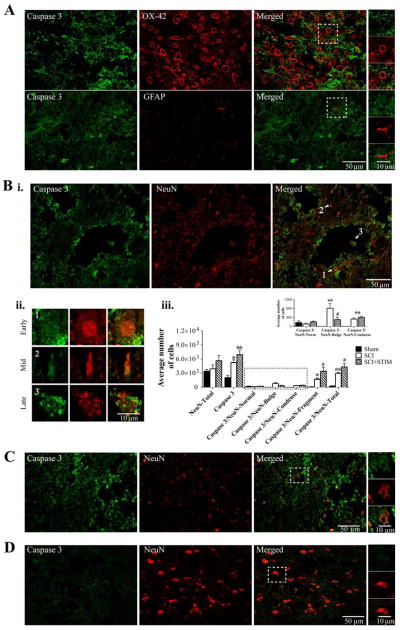
Fig. 11.
Caspase 3 immunofluorescent labeling in the lesioned spinal cord
A) Noxious stimulation increased caspase 3 (green) expression in the lesioned spinal cord. Caspase 3 is co-labeled with OX-42, (red, top panel), but not with GFAP (red, bottom panel). The areas in the white square boxes are enlarged in the panels to the right to further illustrate co-labeling with OX-42 (top) and GFAP (bottom). B) (i) Caspase 3 (green) is also co-labeled with NeuN (red). Extensive caspase 3 labeling is also observed in processes and in areas bordering cavities. (ii) Varying stages of apoptosis (white arrows in Bi, merged image) are demonstrated in neurons. Cells labeled 1, 2, and 3 are enlarged to illustrate (1) a neuron during the early onset of apoptosis, containing a large condensed nucleus, (2) a neuron farther along in the process, containing a shriveled nucleus, and (3) a neuron undergoing late apoptosis, containing a fragmented NeuN-immunopositive nucleus. (iii) Unbiased stereology was used to estimate the average caspase 3 expression in the gray matter of the lesioned epicenter, as well as caspase 3 co-expressed with NeuN depicting the different morphologies (undergoing the varying stages of apoptosis). Caspase 3 expression was greatest in SCI+STIM subjects (p < .01). Caspase was also significantly elevated in SCI subjects (p < .05) compared to sham controls (also see C and D). Caspase 3 was mostly co-expressed with NeuN in the late stage of apoptosis (NeuN-Fragment). Although SCI subjects showed significant caspase 3/NeuN-Fragment co-labeling compared to sham (p < .01), levels in SCI+STIM subjects were significantly greater compared to both SCI (p < .05) and sham groups (p < .001). Caspase 3 expression was less in cells with the NeuN-Bulge and NeuN-Condense morphologies. Yet, caspase 3/NeuN-Bulge and caspase 3-NeuN-Condense co-expressions were significantly greater in the SCI only (p < .01) and SCI+STIM & SCI groups (p < .01), respectively compared to sham. Overall NeuN labeling was comparable across all groups; and neurons with normal morphology, including sham subjects had minimal caspase 3 expression (inset; also see D). [*, indicates significance compared to sham controls and #, indicates significance compared to SCI]. C and D illustrate caspase 3 (green) and NeuN (red) labeling in SCI only and sham subject, respectively. All spinal cord sections are from the mid dorsal spinal cord of the lesioned epicenter.

