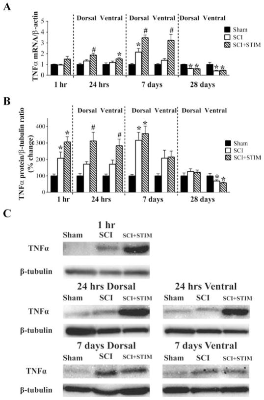
Fig. 3.
TNFα mRNA and protein were increased by SCI and SCI+STIM
A) qRT-PCR analyses showed that neither SCI nor SCI+STIM treatments altered TNFα mRNA levels at 1 hour group after treatment. TNFα mRNA levels in both dorsal and ventral spinal cords were significantly elevated by SCI plus noxious stimulation at 24 hours, post treatment compared to sham surgery. In the dorsal spinal cord the mRNA levels were also significantly increased when compared to SCI alone. Elevated expression of TNFα mRNA in the SCI+STIM was maintained for 7 days, with levels been significantly higher than both sham and SCI group in both dorsal and ventral spinal cord. In the dorsal spinal cord, TNFα mRNA levels in SCI only subjects was significantly increased compared to shams, although levels were still lower than in SCI+STIM subjects. By 28 days, TNFα was decreased in both treatment groups compared to shams. B) At 1 hour and in the dorsal spinal cord at 7 days after treatment, SCI significantly increased TNFα protein levels. Though levels were significantly increased compared to sham, noxious stimulation did not produce any greater effect than SCI only. A significant effect of noxious stimulation was observed in the dorsal and ventral spinal cord at 24 hour. At 28 days, although TNFα levels in the dorsal spinal cord of SCI and SCI+STIM subjects had returned to sham control levels, TNFα levels in the ventral spinal cord were significantly reduced in both treatment groups. [* indicates p < .05 relative to the sham control group, and # indicates p < .05 of the SCI+STIM group relative to the SCI group]. C) Examples of representative western blot images for TNFα. β-tubulin served as a loading control protein.

