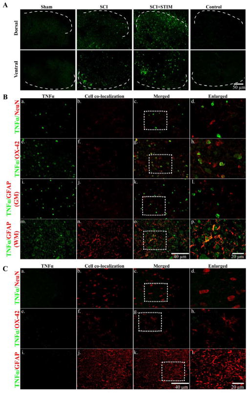
Fig. 4.
TNFα immunofluorescent labeling
A) At 24 hours after treatment, noxious stimulation increased TNFα expression in the lesioned spinal cord (dorsal; top panel and ventral; bottom panel) compared to sham and SCI subjects. The dorsal and ventral spinal cords are devoid of TNFα labeling in tissue from sham controls. In SCI+STIM subjects, TNFα is strongly expressed in the superficial dorsal horn which extends into the deeper laminae of the dorsal horn. TNFα is also strongly expressed in the mid-ventral spinal cord. No labeling was observed on control slides. [Sections depict the dorsal and ventral gray matter. The dotted white lines indicate the gray/white matter border]. B) Further histological characterization of TNFα labeling in the dorsal spinal cord of the lesioned area at 24 hours after stimulation shows that TNFα (green; a, e and i) was co-expressed with the microglial marker, OX-42 (red; f–h). There was no co-labeling of TNFα with NeuN (red; b–d) or GFAP in the gray matter (red; j–l). In the adjacent lateral white matter, TNFα (green; m) is co-localized with GFAP (red; n–p). To further illustrate TNFα co-labeling with NeuN, OX-42 and GFAP, the areas indicated by the white rectangles are enlarged in (d), (h) and (l & p), respectively. C) TNFα expression (green; a, e and i) is negligible in sham control tissue. There is no co-localization of TNFα with NeuN (red; b–d), OX-42 (red; f–h) and GFAP (red; j–l) in sham subjects. Sections in B and C are from the dorsal spinal cord of the lesioned epicenter.

