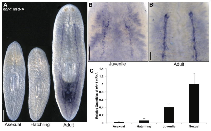
Fig. 2. nhr-1 expression increases with sexual maturation.
(A) Whole-mount, colorimetric in situ hybridization of nhr-1 on asexual, newly hatched sexual, and adult sexual worms. Ventral view shown for all animals. (B–B′) Whole-mount, colorimetric in situ hybridization on juvenile sexual and adult sexual worms to detect nhr-1+ cells in anterior ventral areas. (C) Quantitative PCR showing nhr-1 expression levels at different stages of sexual maturity. Three biological replicates were analyzed for the sexual, asexual, and newly hatched stages. Six biological replicates were analyzed for the juvenile stage due to their variability in sexual maturation. Error bars show 95% confidence intervals. mRNA levels for asexual, hatchling, and juvenile worms are normalized to sexual worms, all differences are statistically significant (p < 0.01, t-test). Scale bars: (A) 500 μm. (B, B′) 200 μm.

