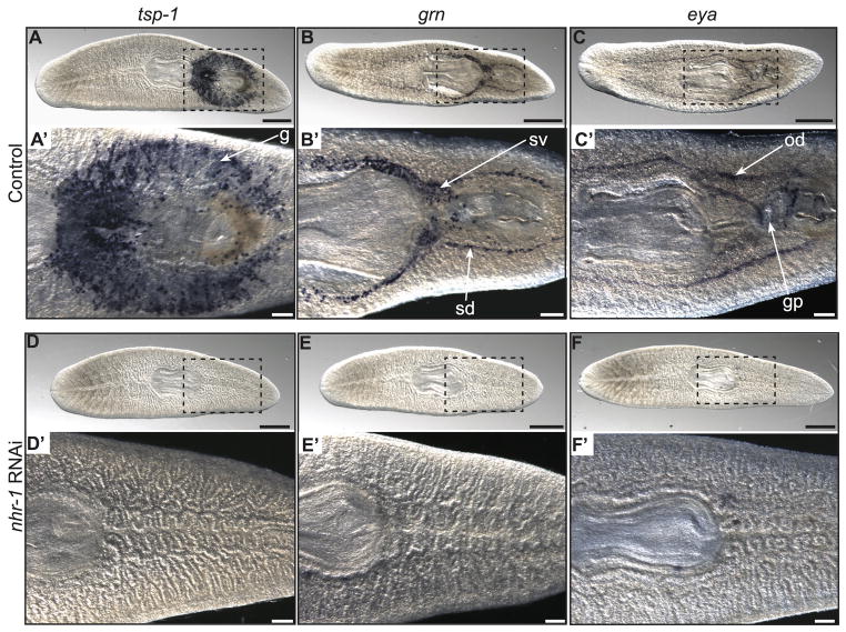
Fig. 3. Disruption of nhr-1 prevents development of accessory reproductive organs.
(A–C′) Whole-mount, colorimetric in situ hybridization to detect tsp-1 in the cement glands (g), grn in the sperm ducts (sd) and seminal vesicles (sv), and eya in the oviducts (od) in adult sexual animals after being fed a control double-stranded RNA. (D–F′) In situ hybridization for tsp-1, grn, and eya after being fed nhr-1 double-stranded RNA. Expression of these genes is not detected. The gonopore (gp) is also missing from nhr-1 knockdown animals. Ventral view shown for all animals. Data are representative of three individual planarians. Scale bars: (A–F) 500 μm. (A′–F′) 200 μm.

