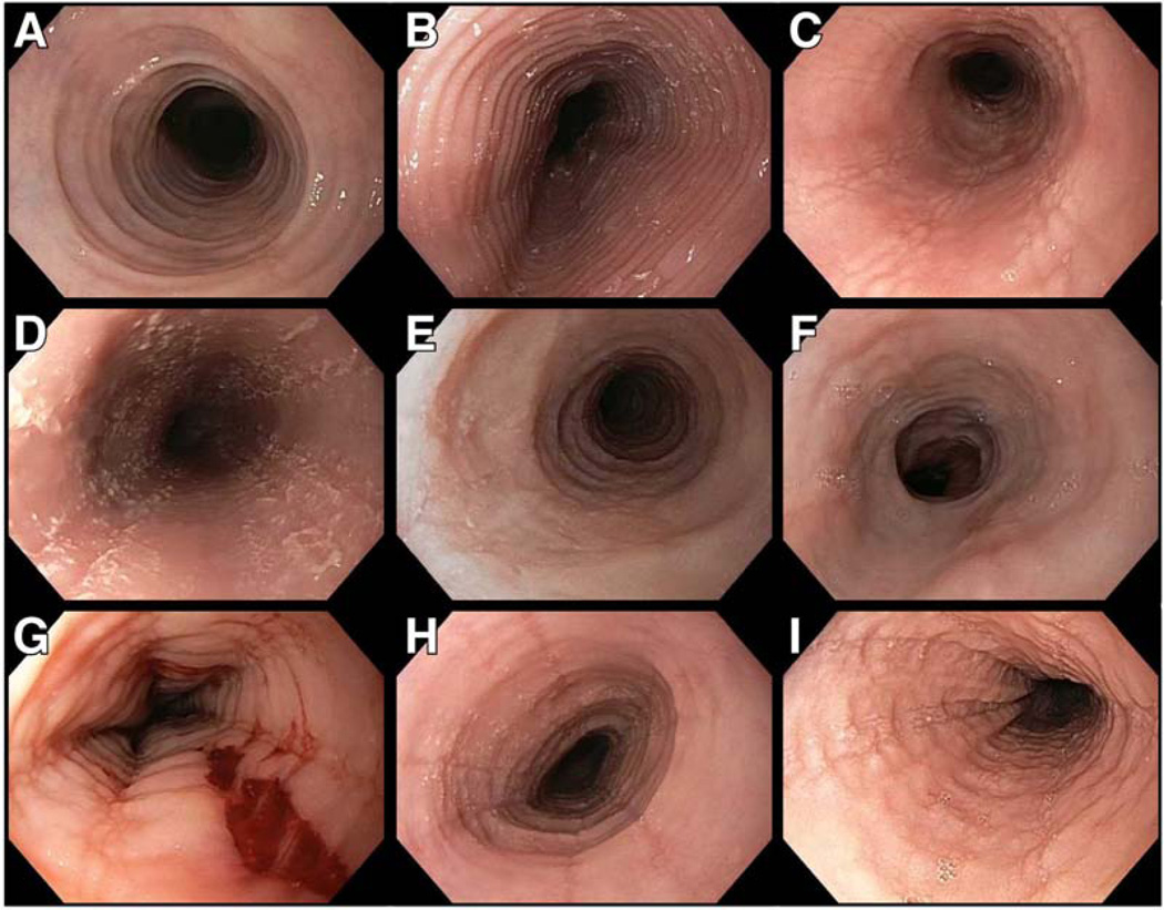
Figure 1.
Endoscopic findings in EoE. (A) Fixed esophageal rings, previously called trachealization. Rings can vary in severity from subtle ridges to tight fibrotic bands, and full insufflation of the esophagus is required to appreciate their extent. (B) Transient esophageal rings, also called felinization. (C) Linear furrows, which are fissures that run parallel to the axis of the esophagus and have a train track appearance. (D) White plaques/exudates, which are eosinophilic micro-abscesses that can be confused with candidal esophagitis; brushings from this patient were negative for candida. (E) Esophageal narrowing with mucosa edema and decreased vascularity. Of note, decreased vascularity and mucosal edema are also visible in images C, D, G, H, and I, and can be a subtle finding in EoE. (F) A more focal stricture in the distal esophagus. Strictures can be located at any location in the esophagus, however. (G) Crêpe-paper mucosa, in which there is a mucosal tear with passage of the endoscope in a narrowed esophagus. (H) A combination of multiple findings including rings, furrows, plaques, narrowing, and decreased vascularity. (I) A combination of several findings including rings, deep furrows, plaques, and mucosa edema.

