2.
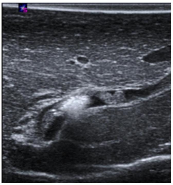
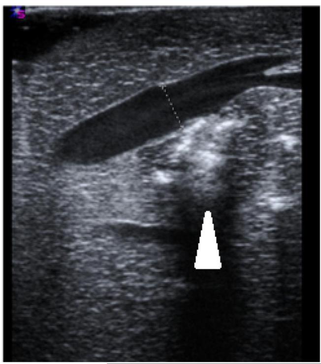
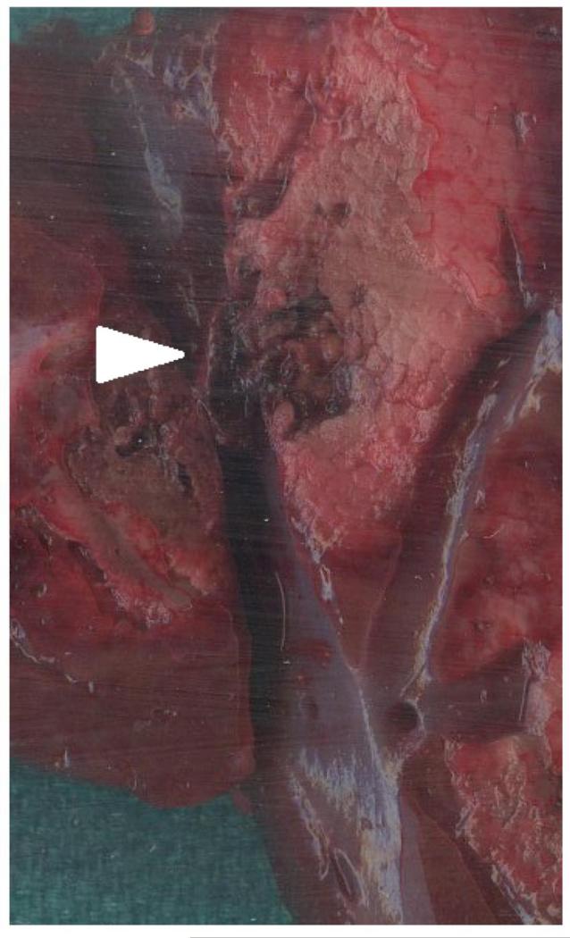
Post-ablation vessel imaging. A) Thrombus formation seen in a nearby portal vein immediately on ultrasound after the ablation procedure at 100 W for 5 minutes. B) Patent hepatic vein immediately after microwave ablation zone creation. Note the absence of thrombus in the hepatic vein, even though the ablation zone abutted the vessel wall, as denoted by the hyper-echoic gas bubbles made from the ablation zone (arrowhead). C) Axial slice of ablation zone with a thrombosed portal vein (arrowhead) inside an ablation zone.


