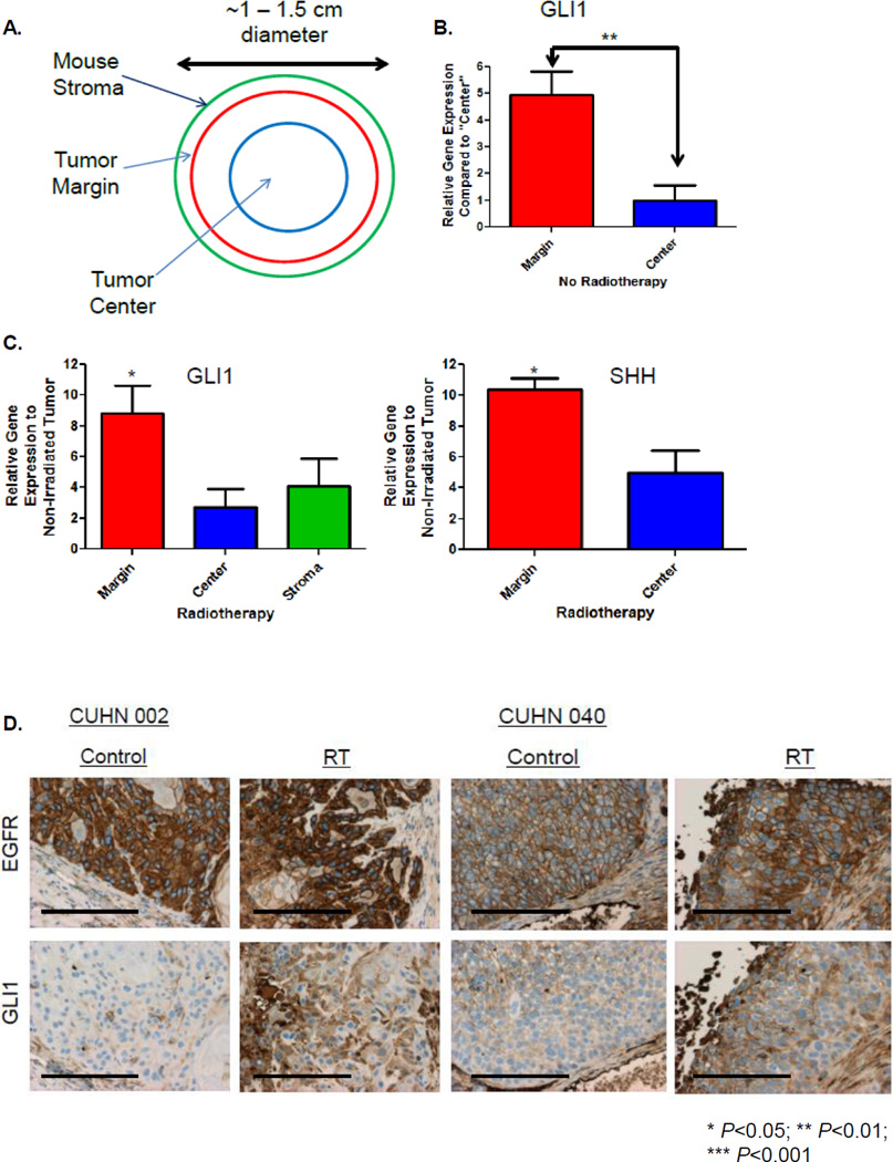
Figure 1. RT Induces GLI1 Expression In Vivo.
A. Schema describing tumor sections of interest that were laser capture microdissected (LCM). B. The amount of non-irradiated human GLI1 mRNA expression at the tumor margin compared to the tumor center was determined by quantitative real-time PCR (qRT-PCR) from LCM tissue. C. Irradiated and non-irradiated LCM tissue was analyzed by qRT-PCR using species specific probes for GLI1 and SHH to determine RT-induced gene fold expression over baseline. D. Two different PDX±RT. Tumors were harvested, formalin fixed, sectioned and stained with anti-EGFR or anti-Gli1 for IHC (20×, bar 100µM). Statistically significant findings were denoted: *P<0.05.

