
Figure 1.
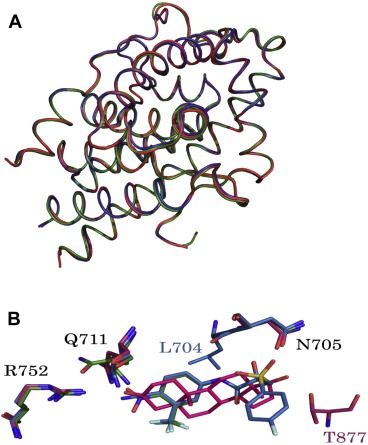
(A). Superposition of the overall structures of wt‐AR‐LBD (red), T877A‐AR‐LBD (green), and W741L (blue)‐AR‐LBD, drawn as ribbon models. (B). Overlapping crucial residues in the active site. Residues are shown as sticks. Nitrogen and oxygen atoms are colored blue and red, respectively. DHT, HF, and CDX are illustrated as red, green, and blue, respectively. Fluorine, nitrogen, oxygen, and phosphorous atoms are depicted as light green, blue, red, and orange, respectively.