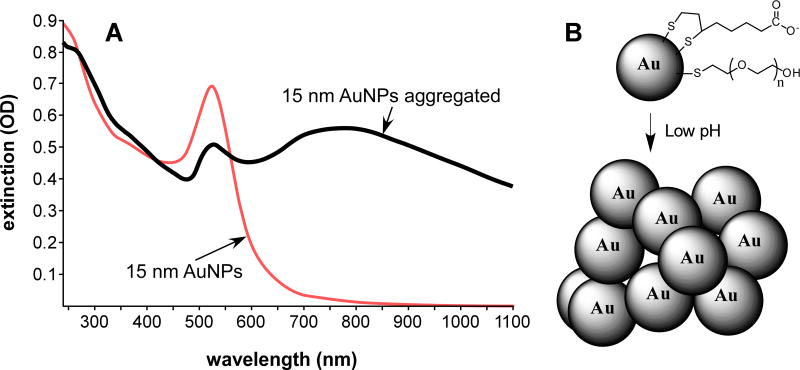
Figure 1.
(A) Change in spectra of 15 nm AuNPs before (red) and after (black) aggregation, here induced by lowering the pH to 5 in 150 mM NaCl. A large increase in the near infrared absorbance (750–1000 nm) is evident. (B) Aggregation of AuNPs at low pH. AuNPs coated with lipoic acid and PEG show aggregation at the pH of endosomes and lysosomes. Gold core size of the AuNPs was measured to be 15.3±1.0 nm by electron microscopy and the hydronamic diameter in solution was measured to be 21.6±0.1 by dynamic light scattering. The thiol ligands are not removed upon aggregation (here reversible with pH), but are not shown in the aggregated clump for clarity.

