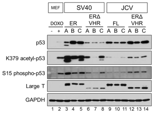
Figure 6. p53 is stabilized and shows different levels of activation in full length and VHR-truncated early region expressing MEFs.
Steady-state levels of total and post-translationally modified p53 protein were detected by western blot in T antigen expressing cells. Large T antigen levels were detected with a combination of SV40- and JCV-specific antibodies (see Methods) and are included for comparison. MEFs treated with doxorubicin were used as a positive control for p53 stabilization and activation. GAPDH, loading control.

