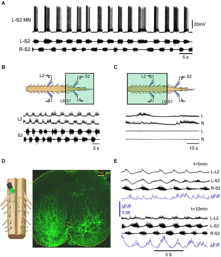
Figure 4.
The methoxamine-induced 0.25–1 Hz rhythm in the sacral spinal segments is relayed to the rostral lumbar network by sub-clusters of ventrally located sacral VF neurons. (A) Intracellular recordings from a left S2 motoneuron (L-S2 MN) are superimposed with concurrent recordings from the left and right S2 ventral roots in the presence of bath-applied methoxamine (100 μM) to the surgically detached sacral segments of the spinal cord. (B) Recordings from the left and right L2 and S2 ventral roots showing alternating left-right bursting when methoxamine is applied to the sacral compartment (colored rectangle) of a dual chamber experimental bath. (C) Recordings from the left and right L2 and S2 ventral roots in the experiment described in (B) after washing the sacrally applied methoxamine and adding methoxamine to the TL compartment (colored rectangle) of the experimental bath. The sacral rhythmic activity is blocked, while a slow rhythm appears in the lumbar segments. (D) Confocal micrograph of a 70 μm cross section through the S2 spinal segment shows left sacral neurons back-loaded with fluorescein-dextran through the right VF at the lumbosacral junction (dye back-loading is illustrated on the left). (E) Imaging the activity developed in a ventral S2 VF-neuron back-loaded with Calcium green dextran and viewed from the ventral aspect of the isolated spinal cord (blue, ΔF/F) and the concurrently recorded motor output from the left L2 and the left and right S2 ventral roots, 5 and 10 min after addition of methoxamine to the experimental bath. The 0.1 Hz–10 Khz ventral root recordings were not high-pass filtered to reveal the early subthreshold activity in L–L2.

