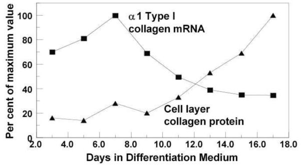
Fig. 2. Relationship between type I collagen mRNA and cell layer collagen protein accumulation.

MC3T3-E1 osteoblastic cell line was cultured in differentiation medium, and gene expression of Col1a1 and cell layer type I collagen contents were analyzed by Northern blot and amino acid analysis, respectively. Col1a1 gene expression was highest at day 7 and decreased gradually thereafter, while extracellular collagen accumulation became evident after 9 days of culture. Such discrepancies occurs, in part, due to the complex biosynthesis process including post-translational modifications (shown in Fig.1) Modified from (Hong et al., 2004)[53].
