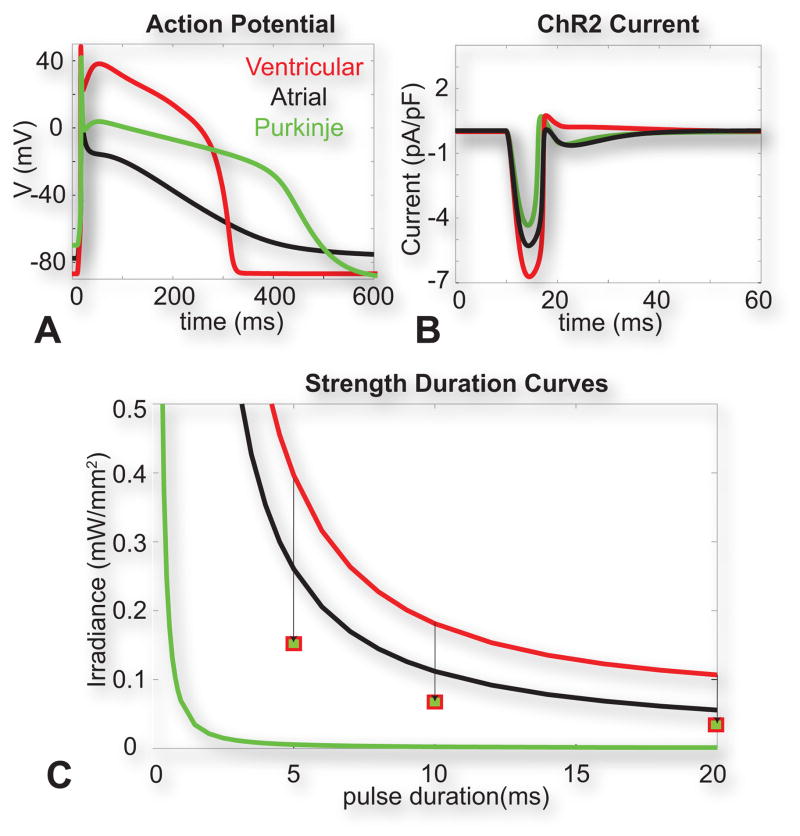
Figure 3. Optical excitation in human cardiac cell types.
A. Optically-triggered action potentials (10 ms pulse at 470nm, 0.5 mW/mm2) in human ventricular, atrial and Purkinje cells. B. Underlying ChR2 current upon the action potential generation for the three cell types. C. Strength-duration curves for the three cell types. Squares show simulated values for optical excitation threshold in ventricular myocytes with a Purkinje formulation of IK1. Reproduced with permission from (Williams et al., 2013).

