
Figure 7. In vitro and in vivo cardiac expression of AAV1.
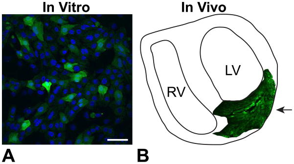
A. Neonatal rat ventricular cardiomyocytes infected with AAV1 in vitro at MOI 1000. Green fluorescence indicates eGFP reporter and blue indicates DAPI nuclear staining. Scale bar is 50 μm. B. Schematic of a slice of an adult rat heart infected with 1×10¹¹ AAV1 particles, delivered via intramyocardial injection 4 weeks earlier, as indicated by the arrow.