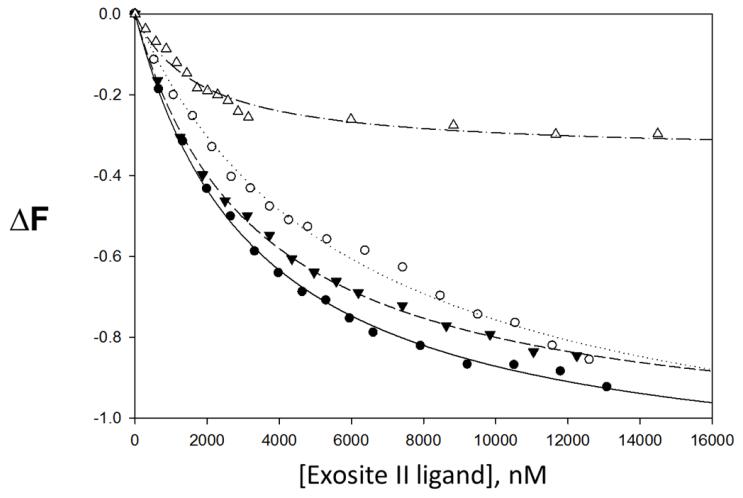
Figure 4.
Change in thrombin↔p-aminobenzamidine (PABA) complex fluorescence with increasing concentrations of exosite II ligand. • CDSO3, ○ FDSO3, ▼ SDSO3, △ heparin. ΔF is the relative change in fluorescence. Line of best fit for CDSO3 (—), FDSO3 (----), SDSO3 (---) and heparin (-----). PABA reversibly binds to the active site of thrombin.

