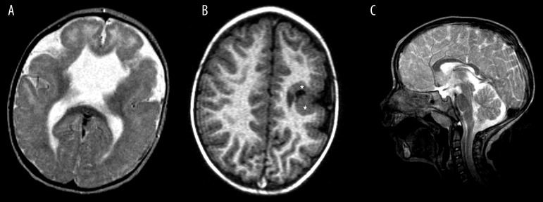
Figure 4.
MRI features of the common abnormalities associated with schizencephaly. (A) Agenesis of septum pellucidum – axial T2-weighted image. (B) Gray matter heterotopy in the vicinity of schizencephalic cleft (stars)– axial plane T1-weighted image. (C) Dysgenesis of the corpus callosum (arrow) – sagittal plane, T2-weighted image.

