
Figure 6.
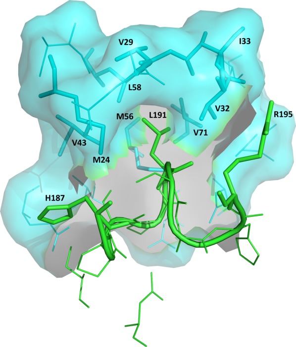
Binding of UNG to Ugi. Close-up view of the protein-Ugi interactions in the eUNG-Ugi complex [1UUG]. The figure emphasizes the insertion of Leu191 in the Leu-intercalation loop (residues 187–195 shown as cartoon in green) of eUNG into the hydrophobic cavity of Ugi (shown as surface in cyan). Residues His187, Leu191, and Arg195 of the Leu–intercalation loop in UNG and residues Met24, Val29, Val32, Ile33, Val43, Met56, Leu58, and Val71 in the hydrophobic cavity of Ugi are represented as stick models. These 11 residues are labeled. The remaining residues in this close-up are shown as thin lines (Ugi in cyan; UNG in green). An interactive view is available in the electronic version of the article.