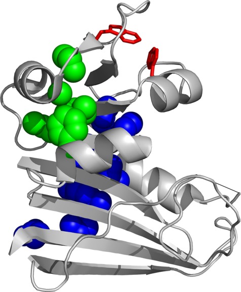
Figure 1.

Structure of DHFR. The ribbon representation of the structure of DHFR (PDB CODE: 5DFR) is shown. The side chains of the residues in the two hydrophobic clusters that form in IBP are shown in blue and green spheres. The side chains of Trp47 and Trp74, which are responsible for high fluorescence in IHF, are shown in red sticks. The invisible part of the Met20 loop is shown in a straight line. The image was created with PyMOL.
