
Figure 3.
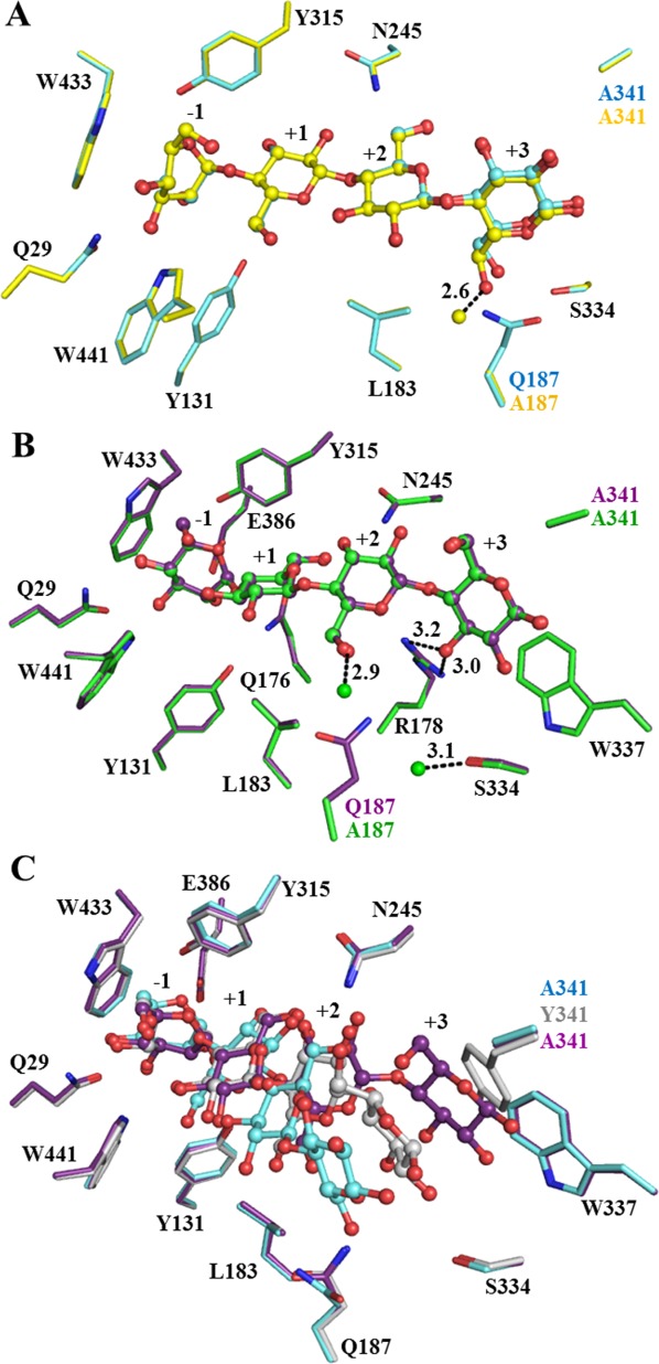
Cellotetraose binding in the active site of BGlu1 E386G/Y341A/Q187A, BGlu1 E176Q/Y341A and BGlu1 E176Q/Y341A/Q187A. (A) Comparison of cellotetraose binding in active sites of BGlu1 E386G/Y341A (cyan carbons) and BGlu1 E386G/Y341A/Q187A (yellow carbons). The fixed water molecule seen in E386G/Y341A/Q187A, but not in E386G/Y341A is shown as a yellow ball. (B) Cellotetraose binding in the active sites of BGlu1 E176Q/Y341A (violet carbons) and BGlu1 E176Q/Y341A/Q187A (green carbons), which has new fixed water molecules that are shown as green balls. In both BGlu1 E176Q/Y341A and E176Q/Y341A/Q187A, O3 of Glc4 forms direct hydrogen bonds with R178 (shown as dashed lines) and Glc4 was stacked upon W337. (C) Cellotetraose in the active sites of BGlu1 E176Q (gray carbons), BGlu1 E386G/Y341A (cyan carbons) and BGlu1 E176Q/Y341A (violet carbons). The cellotetraose is represented as balls and sticks.